
What to Expect From a 3D Mammogram
The 3D mammogram procedure provides new technology providing an array of benefits over standard mammography. Many medical professionals recommend you have a mammogram beginning at age 40 if you have an average risk of breast cancer. But, the American Society of Breast Surgeons has taken this one step further. They released an updated position statement which states women with an average risk of developing breast cancer should preferably have a 3D diagnostic mammogram performed yearly.
What Is a 3D Mammogram?
Also known as breast tomosynthesis, 3D mammography is a relatively new procedure for breast imaging that the FDA approved in 2011. Like traditional mammograms, 3D mammography uses x-rays to produce breast images to detect tumors, lumps or other abnormalities. It can produce breast tissue images in more detail.
Having routine mammograms provides you with several benefits, including:
- Providing you with a 30% reduced risk of dying from breast cancer
- Saving your life by detecting breast cancer early
- Allowing you to receive early treatment, which means there is a greater chance of you keeping your breasts without having to undergo mastectomy
3D captures multiple slices of your breast at various angles. Then, the doctor brings the images together to create an incredibly clear 3D reconstruction of your breasts to review one slide at a time, just like turning a book’s pages. This makes it simpler for your doctor to see if there’s a need for concern.
The 3D representation of your breast is similar to CT technology created 3D images. Tomosynthesis is different from CT technology in that it projects substantially fewer x-ray beams through your breast than CT technology does, and there’s dramatically less x-ray exposure to the rest of your chest.
Although 3D mammography uses extremely low-dose x-rays, it is currently often used often along with 2D mammography, which makes the total radiation dose higher than there would be with just standard mammography. Early assessments of tomosynthesis suggest an enhanced detection of breast cancer than seen with 2D mammograms, but substantial large-scale comparisons of 3D mammography with 2D mammography in studies are still in process.
What Are the Risks of a 3D Mammogram?
Since a mammogram uses x-rays for producing breast images, you have exposure to small amounts of ionizing radiation. The benefits of mammography, for most women, outweigh the risks this small amount of radiation poses. The risk linked with this dose seems to be greater in women under the age of 40. But, in some cases, mammography’s benefits for detecting breast cancer under 40 years old still outweigh the risk of radiation exposure.
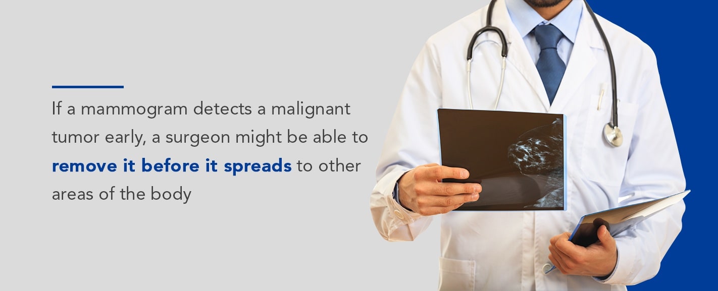
For instance, mammography might reveal that a mass is benign, and therefore won’t require treatment. Also, if a mammogram exam detects a malignant tumor early, a surgeon might be able to remove it before it spreads to other areas of the body, which would require more aggressive treatment like radiation or chemotherapy.
Still, there are some risks to consider, including:
- Low radiation level exposure. As mentioned, you have exposure to small amounts of radiation with 3D mammography, and since it is usually combined with traditional mammography, you’re exposed to even more radiation than you would be with the traditional mammogram alone. But, there are now some newer 3D mammogram devices that can generate both 2D and 3D images together, which lowers your radiation exposure.
- A 3D mammogram might detect something suspicious that turns out to not be cancerous. A 3D mammogram could detect an abnormality that appears to be consistent with normal tissue or benign after further testing. This is what doctors call a “false positive” result, and it could cause you unnecessary anxiety when undergoing additional testing and imaging like a biopsy to evaluate the suspicious area further.
- 3D mammogram can’t detect all cancers. There is the possibility the 3D mammogram process could miss an area of cancer, like a very small cancer or an area that’s hard to see.
Why Is a 3D Mammogram Performed?
Doctors use a 3D mammogram as a breast cancer screening tool to screen those with no symptoms or signs of breast cancer. They also use it to evaluate breast problems like thickening or a suspicious lump. When it’s used for breast cancer screening, it creates both 3D and standard 2D mammogram images since both image types have some benefits in seeing certain breast abnormalities.
Combining a 3D and standard 2D mammogram can:
- Detect slightly more cancers than just operating a 2D standard mammogram alone would. Research shows combining the two types of mammograms can result in around one more breast cancer for every 1,000 females screened detected when compared with 2D mammography alone.
- Decrease the need for follow-up imaging. When physicians find abnormalities on traditional mammogram images, they might suggest additional imaging. It can be stressful having to come back for additional imaging. It might take extra time, leading to extra costs. 3D mammograms help radiologists distinguish between benign structures and tumors, which may prompt fewer call-backs for false positives. This, in turn, reduces the overall anxiety, stress or fear for patients.
- Enhance the detection of breast cancer in dense breast tissue. 3D mammography offers benefits in breast cancer detection in individuals with dense breast tissue since the 3D image allows physicians to see beyond the dense areas.
Breast tissue consists of milk ducts, milk glands and dense or supportive breast tissue and fatty tissue. Dense breasts consist of more dense breast tissue than fatty tissue. The breast tissue and cancer both show up white on a traditional mammogram, making breast cancer harder to detect in dense breasts.
There’s not enough evidence for concluding 3D mammography could reduce the risk of dying due to breast cancer more than traditional mammography alone. Because of this, most breast cancer screening guidelines don’t specify women should opt for 3D mammograms over traditional mammograms alone.
How to Prepare for a 3D Mammogram
To help prepare for your mammogram, follow these tips:
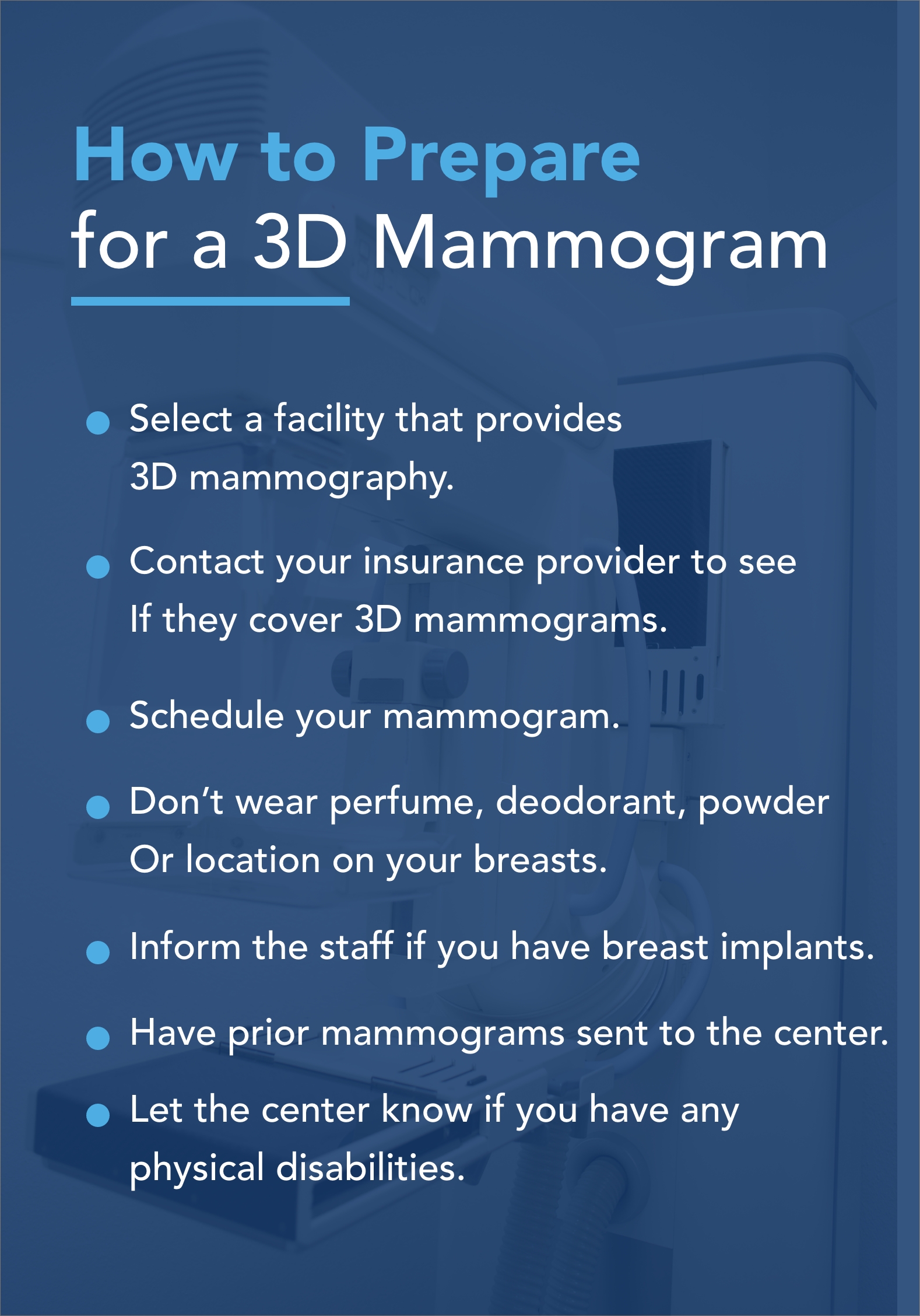
- Select a facility that provides 3D mammography. Even though 3D mammography is becoming more common, they’re still not available everywhere. Your doctor should be able to help you find a place in your area where 3D mammography is available.
- Contact your insurance provider to see if they cover 3D mammograms. Not all insurers do. Contact them before your test so you know what type of out-of-pocket costs you should expect. They might cover the traditional mammogram part of the test, and you’d be responsible for the 3D mammogram part cost.
- Schedule your mammogram during a time you think your breast will be the least likely to be tender. This is typically during the week following your menstrual period if you haven’t gone through menopause yet. It’s likely your breasts will be the most tender the week before your period and the week during it.
- Don’t wear perfume, deodorant, powder or lotion on your breasts or under your arms on the day of your screening. It could cause foreign particles to show up on the images.
- Inform the staff if you have breast implants. They might have to take additional images than a traditional mammogram.
- Have prior mammograms sent to the center. Or, if you have copies, bring them with you.
- Let the center know if you have any physical disabilities. These could make it difficult for you to lift your arms, sit up or hold your breath.
What Can You Expect During a 3D Mammogram?
How is a 3D mammogram done? Here’s what to expect during your 3D mammogram.
At the testing center, your technologist will provide you with a gown to wear, and ask you to remove all clothing and jewelry from the waist up. Wear a two-piece outfit the day of your test to make this easier.
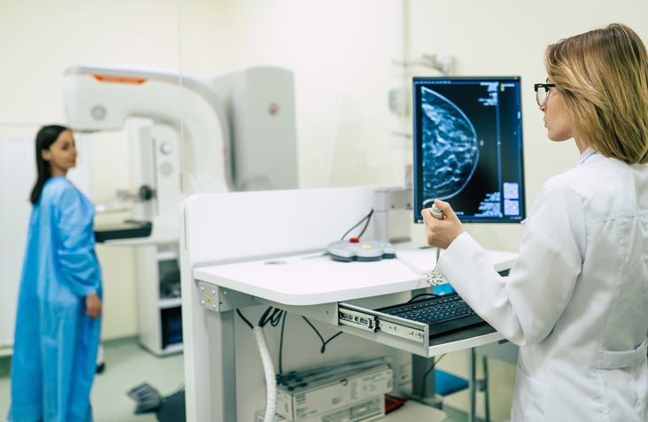
For the test itself, you’ll stand in front of the x-ray machine equipped for performing 3D mammography. The technologist will place one of your breasts on a platform, raising or lowering the platform to match your height. They’ll help you position your arms, head and torso to allow an unobstructed image of your breast.
A clear plastic plate gradually presses your breast against the platform. It applies pressure for several seconds to spread your breast tissue out. While this pressure isn’t harmful, it could be uncomfortable and even painful. Let the technologist know if you’re having too much discomfort.
The 3D mammogram device moves from one side to the other above you to collect images. You might need to stand still and hold your breath for several seconds to minimize movement.
The machine releases the pressure on your breast, and the technologist repositions the machine to take a side view image of your breast. They’ll position your breast on the platform again and the plastic plate will once again apply pressure. The technologist takes images and repeats the process on your other breast.
How Is a 3D Mammogram Different From a 2D Mammogram?
Standard digital mammograms take two-dimensional images of your breast and are still one of the most innovative tools for detecting abnormalities in breasts. However, instead of viewing 2D images of breast tissue, doctors can now view the tissue one layer at a time — think flipping pages of a book. The doctor can view more fine details since it will be less likely that overlapping tissue will hide them.
Let’s look at the primary differences between 2D and 3D mammography.
1. The Mammogram Procedure
While the physical discomfort you might experience is no different between the two, the 3D mammogram screening does take several seconds longer.
2. Availability of 2D and 3D Mammograms
The 3D mammograms are newer than 2D mammograms and might not be available at all facilities. Plus, 3D mammograms could cost a little more. Some insurance providers won’t cover 3D mammograms.
3. Accuracy of 2D and 3D Mammograms
Since the radiologist can scroll through the breast tissue images one layer at a time with 3D mammography, it’s easier to identify abnormalities. And, since the 3D images provide more detail, they decrease the false-positive rate. Therefore, it’s less likely you’ll need to go back for another mammogram or require more testing. This can help cut down anxiety and stress.
3D mammograms have a slightly higher rate of cancer detection than 2D mammograms. A study that involved several years of data found the 3D mammogram’s benefits last over time.
If your doctor has told you your breast tissue is dense, it means you have more supportive tissue in your breasts than fatty tissues. Supportive tissue shows up white on mammograms, but cancer does too. 3D mammograms make it simpler for the radiologist to tell the difference between cancer and supportive tissue, reducing the chances of a false-positive result, which would require more testing.
4. Radiation from Mammograms
Clinical data reveals there is a small increase in the amount of radiation with 3D mammography involves over 2D mammography. Medical research indicates that a small dose increase of radiation should not deter the use of 3D mammograms. And, 3D mammograms enable doctors to see beyond areas of density in women with dense breast tissue.
What to Expect From a 3D Mammogram
The images gathered during 3D mammography are synthesized by a computer to form the 3D image of your breast. The doctor can analyze the images the 3D mammogram produces as a whole or in small segments for greater detail. For breast cancer screening, the machine creates 2D mammogram images as well.
The radiologist evaluates the images to check for any abnormalities potentially indicating breast cancer. If they see anything abnormal, they’ll use the 2D mammogram images to determine if additional testing is needed.
Additional breast cancer tests might include an MRI, ultrasound or, in some cases, a biopsy for removing suspicious cells to test in a lab by physicians specializing in analyzing body tissue.
Benefits of Choosing Clear Connect Medical Imaging for Your 3D Mammogram
Clear Connect Medical Imaging maintains the foundation of consistency and efficiency while revolutionizing imaging. We’re committed to outstanding and high-quality service and combine that with innovative technology with a focus on exceptional patient care. We’re proactive about technological innovation and advancement, so you’ll receive the most comprehensive, modern services.
We combine this stellar service with a dedicated attentiveness to your personal needs, which makes us your premier medical imaging location. Benefits of choosing Clear Connect Medical Imaging include:
- Friendly staff: Our staff is dedicated to being friendly, timely and helpful.
- Individualized patient care: Our imaging experts care about every patient as an individual and whole person. You’re treated with the respect you deserve.
- Advanced technology: We’re constantly evolving in our imaging technology and understanding of the industry. Clear Connect Medical Imaging is proud to offer 3D mammograms.
- Comfort: We deliver our services in light-filled, elegant and comfortable offices.
- Compassion: We show compassion by accommodating your needs and providing you with a pleasant and accurate imaging experience.
![]()
Schedule an Appointment With Clear Connect Medical Imaging for Your Breast Cancer Screening
We invite you to visit any of our locations to experience our hospitality and professionalism. You’ll learn why Clear Connect Medical Imaging is an industry leader in quality care and patient satisfaction.
The staff at Clear Connect Medical Imaging is specialized in helping you with your medical imaging needs.
To schedule an appointment for your 3D mammography, contact Clear Connect Medical Imaging today.
