

Often, doctors need to look beneath the surface to get a clear idea of what their patient may be experiencing. That’s where penetrative imaging is valuable.
Let’s explore what penetrative imaging is, how it works and when you might need it.
Jump Ahead:
How does penetrative imaging work?
Penetrative imaging produces black-and-white images doctors can use to examine internal structures for easier diagnosis. Depending on the type of test, the imaging process may use radiation or magnetism to capture the picture.
Sometimes, an imaging exam involves contrast media to produce a clearer image of the body parts under examination. This substance is a dye that increases the contrast between different parts, which helps radiologists make more accurate diagnoses. Technologists often administer contrast in one of three ways:
- Oral liquid
- Injection
- Enema
Each type of imaging has different purposes and benefits.
3 types of penetrative imaging
Doctors can choose from many different types of penetrative imaging to provide a glimpse into the body. Here’s a quick overview of today’s three most common imaging modalities.
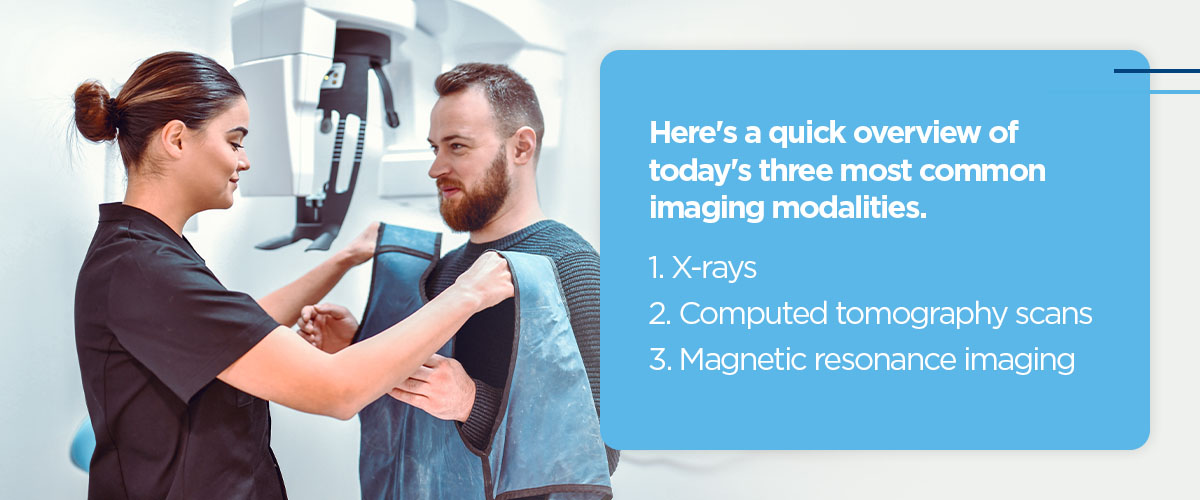
1. X-rays
Most people are familiar with x-rays as the oldest and simplest type of penetrative imaging. X-rays use beams of ionizing radiation to produce images of internal structures — especially bones. Here’s how it works.
- Positioning: You will lie, sit or stand on the x-ray table and angle yourself to provide the best view of the body part you are getting. The technologist may cover other parts of your body with a lead apron to minimize radiation exposure.
- Exposure: The technologist will focus the x-ray beam on the body part and step behind a protective window before capturing an image.
- Repeat: You may need to reposition yourself so the technologist can get a more complete view.
The best-known application for x-rays is assessing bone conditions like fractures and osteoporosis. That’s because bones and other solid structures block x-ray beams from completely passing through the body, which leaves behind bright white shapes that contrast with gray soft tissues like fat, blood and muscle. In mammograms, x-rays can also help health providers detect and diagnose breast abnormalities.
While you may worry about radiation exposure during x-rays, we assure you the technician will always use the smallest dose possible to capture the images.
2. Computed tomography scans
Also known as computer-assisted tomography, a CT scan is an x-ray technique that produces highly detailed cross-sections, or “slices,” of the area under examination. Our advanced CT scanners at Clear Connect Imaging create “spiral slices,” which produce more complete images of affected tissues in less time than traditional CT machines.
Though CT scanners use x-ray technology, the images typically contain more data thanks to the rotating scanner. Here’s how a CT scan usually works:
- You will lie on the x-ray table, which slides into a round chamber.
- The machine will rotate as you pass through the chamber, capturing x-ray images at various angles. This step usually takes anywhere from 10 to 30 minutes, depending on whether your exam requires the use of contrast.
- Upon completion of the scan, you will be free to go about the rest of your day as usual.
Because CT scans don’t take much time, they’re excellent for use in emergency departments, where physicians need to quickly diagnose life-threatening conditions for effective treatment.
3. Magnetic resonance imaging
An MRI is a noninvasive imaging test that examines soft tissues, like your brain or muscles. Unlike x-rays and CT scans, MRI scans do not use ionizing radiation. Instead, they combine magnetism and radio frequencies to alter atomic movement within the body.
Here’s what the typical MRI process looks like:
- Like in a CT scan, you lie on the examination table, which slides into the MRI scanner. You must remain completely still to ensure the scanner can get a good image. The operator will walk you through the entire exam via intercom.
- The machine will generate a magnetic field that aligns the atoms in your body in the same direction. You may hear loud banging and humming noises during this time, but don’t worry — that’s just the magnetic field.
- Radio waves from the scanner will pass through your body, changing the spin of the atoms.
- When the radio waves shut off, the atoms return to their original alignment. This shift sends radio signals back to the computer, which converts them into a 3D image of the target area.
- Once the exam ends, you can go back to the rest of your day.
The exam can take up to an hour to complete, though it might last slightly longer if you need contrast.
Because the machine uses strong magnets, patients with metal or electronic devices such as pacemakers and cochlear implants may need to explore alternative options. Pregnant patients should also speak with a doctor before receiving an MRI to minimize risk.
Applications and benefits of penetrative imaging
Penetrative imaging enables doctors to assess internal structures, diagnose illnesses and injuries and determine how far a condition has progressed. As a result, these tests enable more accurate diagnoses and effective treatments, which help physicians provide the best care possible.
Here are the most common ways medical providers use these exams.
- X-rays: Mammograms and bone examinations are daily applications for traditional x-rays, but the technology can also aid in diagnosing some infections and issues in the digestive tract.
- CT scans: Typical applications for CT scans include assessing traumatic injuries, detecting cancers, diagnosing heart and vascular disease and even guiding biopsies.
- MRI scans: An MRI scan enables doctors to examine abnormalities in soft tissues, such as aneurysms, tumors, spinal cord disorders, blood clots, joint issues and more.
Learn more about penetrative imaging services from Clear Connect Imaging
If you or someone you love needs medical imaging services, consider asking your physician to refer you to one of our local facilities. Our commitment to cutting-edge technology and compassionate care helps us provide excellent imaging services for patients in every stage of life.
You can learn more about our penetrative imaging services on our website. Questions? Feel free to read our FAQ page to see if we have the answer.
Ready to request your next diagnostic imaging appointment? View our Tallahassee location.